Should ground-glass nodules be examined with PET-CT?
In today's medical field, with the rapid development of imaging technology, there are more diagnostic options available for diseases.
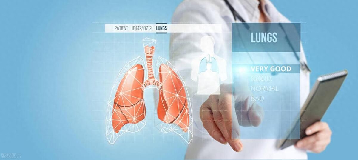
Among them, PET-CT, as an advanced imaging device, is considered an important tool for tumor detection due to its ability to provide both anatomical structure and functional metabolic information.
So, if a ground-glass nodule appears in the lung, is a PET-CT scan necessary?
As a common finding in pulmonary imaging, ground-glass nodules typically refer to hazy, slightly dense, cloud-like shadows with indistinct borders in the lung.
The appearance of such nodules may indicate lung inflammation, hemorrhage, fibrosis, or even early-stage lung cancer.
Therefore, timely and accurate diagnosis is crucial for patients with detected ground-glass nodules.
PET-CT, as the pinnacle of medical imaging technology, achieves tumor localization and qualitative diagnosis by injecting contrast agents containing radioactive isotopes and utilizing the high glucose metabolism characteristics of tumor cells.
This technology not only assists doctors in accurately determining tumor location but also evaluates tumor metabolic activity, providing crucial evidence for treatment planning.
However, PET-CT is not a universal golden key.
For pulmonary ground-glass nodules, especially those smaller than 1cm, PET-CT demonstrates limited diagnostic value.
The reason is that even in early-stage tumors such as carcinoma in situ or minimally invasive carcinoma, the number of tumor cells is limited, and their metabolic activity is relatively low.
Therefore, when the sugar-based contrast agent enters the body, these tumor cells absorb insufficient sugar to produce a noticeable metabolic signal, often resulting in PET-CT scans being unable to accurately determine whether the nodule is benign or malignant.
Thus, when faced with ground-glass nodules in the lungs, patients should remain calm and avoid blindly pursuing advanced diagnostic methods.
For micro-nodules smaller than 1 cm, a strategy of follow-up observation is generally recommended, monitoring changes in the nodules through regular CT scans.
If the nodule shows malignant features such as enlargement, morphological changes, or increased density, further diagnosis and treatment should then be considered.
Additionally, patients can combine other diagnostic methods, such as blood tumor marker tests, bronchoscopy, or needle biopsy, to enhance diagnostic accuracy and reliability.
Each of these examination methods has its own advantages and can provide physicians with diagnostic evidence from different perspectives.