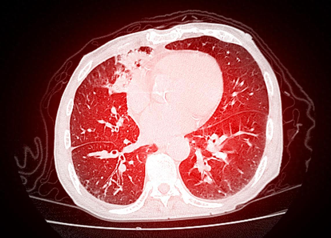
Understanding Your Lung CT Report! Don't Panic When Seeing These Terms
Does receiving a lung CT report full of dense medical terminology make you nervous? Actually, many indicators turn out to be "false alarms"! Master these 11 common descriptions to easily assess your lung health status:
1. Well-defined borders: Lesions with regular contours often indicate benign conditions, requiring no excessive concern;
2. Blurred border: Requires attention but no need for panic; follow doctor's orders for regular follow-up examinations;
3. Fibrotic focus: "Scar" left by lung self-repair, mostly old lesions with high stability;
4. Calcification: Similar to human "stones", commonly seen after inflammation or tuberculosis healing, generally harmless;
5. Linear shadow: "Healing mark" after lung infection, like skin scabbing, usually requiring no special treatment;
6. Small pulmonary nodule (<10mm): Mostly benign hyperplasia, some require regular monitoring;
7. Very small nodule (<5mm): Extremely low probability of malignancy, observation is sufficient;
8. Miliary pulmonary nodules (1-2mm): Multiple small nodules are usually benign reactions, follow-up examinations are recommended;
9. Pleural thickening: A "sequela" after recovery from pleural inflammation, mostly asymptomatic and requiring no treatment;
10. Fibrotic nodule: If present long-term, it's usually chronic inflammation; newly detected cases should be investigated for potential tuberculosis.
11. Increased lung markings: Commonly seen in pulmonary inflammation, long-term smoking, or air pollution. Can be alleviated by improving lifestyle habits.
CT reports serve as a "barometer" of health, but not all abnormalities indicate disease. If you encounter the above findings, don't panic. Consult a doctor promptly and follow up regularly when necessary. Scientific management brings greater peace of mind!