Things look bent? Don’t ignore it! “Visual distortion” may hide serious fundus disease
Let's start with a small test:
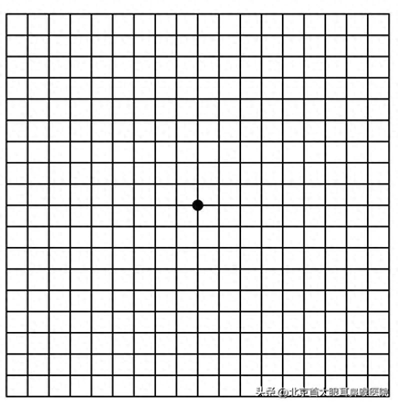
Cover one eye with your hand and focus with the other eye on the black dot in the center of the image below, or on straight-line objects such as window frames or door frames.
Ask yourself, are the lines you see straight and clear? Is any part appearing bent, distorted, missing, or wavy?
(Please imagine the image below as an Amsler grid)
。
If the images you see include portions of lines that appear bent, twisted, or have blurred gaps (as illustrated by the wavy line above), you need to be highly alert! This phenomenon is medically called "metamorphopsia."
A normal retina is flat and smooth, able to accurately receive external images. But when the macular area undergoes disease, becoming edematous, elevated, or pulled, the image projected onto it will become warped and distorted, like looking at a picture printed on an uneven water surface.
Common sensations include:
Straight lines appearing wavy
Door frames appear bent
Objects appear larger or smaller (macropsia/micropsia)
Dark shadows or scotomas in the central visual field
Causes of metamorphopsia are usually relatively serious and mainly centered on the macular region. The following are several of the most common causes:
1. Central serous chorioretinopathy
Young and middle-aged men, under high stress
What happens: choroidal blood vessels under the retina leak, fluid accumulates in the macular area, causing the retina to bulge, which leads to visual distortion and micropsia, accompanied by decreased central vision and duller colors.
Characteristic: has some capacity for spontaneous recovery but prone to recurrence; requires close observation.
2. Age-related macular degeneration
People over 50 years old
What it is: divided into dry and wet types. Wet macular degeneration is the main cause of severe vision loss in the elderly; its essence is the growth of abnormal neovascularization in the macular area. These vessels readily leak and bleed, rapidly destroying macular structure and causing severe metamorphopsia and central vision loss.
Characteristics: progresses rapidly and requires urgent treatment.
3. Epiretinal membrane (macular pucker)
Middle-aged and elderly people, history of ocular surgery or inflammation
What happens: A layer of transparent fibrous cellular membrane grows and contracts on the macular surface, like a wrinkled plastic wrap covering the "negative," pulling on the retina and causing distortion.
Characteristics: The disease progresses slowly and may be asymptomatic in the early stages.
4. Macular hole
More common in elderly and females
What it is: A full-thickness hole forms in the tissue of the central macular area; when looking, a persistent dark spot appears in the center and the surrounding image is distorted.
Features: Usually requires surgical intervention.
5. Pathologic myopia
High myopia (greater than 6.00 D)
What happens: The eyeball in high myopia resembles an overinflated balloon; the globe wall becomes thin and fragile, and the macular area is particularly prone to atrophy, hemorrhage, neovascularization, and other lesions, thereby causing metamorphopsia.
Feature: Patients with high myopia must have regular fundus examinations!
6. Diabetic macular edema
Diabetic patients
What’s happening: Long-term hyperglycemia damages retinal blood vessels, causing vascular leakage; fluid accumulates in the macular area, leading to edema and resulting in blurred and distorted vision.
Features: One of the most common causes of vision impairment in diabetic patients. It may delay the optimal timing for treatment.
This article is for medical popular science purposes only and cannot replace professional medical diagnosis and advice. If you have related symptoms, please promptly visit the ophthalmology department of a reputable hospital.