COVID-19 Led to Lung Cancer Detection? What Tests Should Be Done for Lung Cancer?
The COVID-19 pandemic has been a significant blow to the entire world. For us, it has been both a lesson and a warning. Beyond this, a small number of people have had their lives saved due to the pandemic. Why is that?
I. Lung Cancer Discovered Through Pandemic Screening
The most obvious manifestations of COVID-19 are fever, fatigue, and dry cough. Additionally, there is a highly distinctive imaging feature: multiple ground-glass opacities and infiltrates in the lungs.
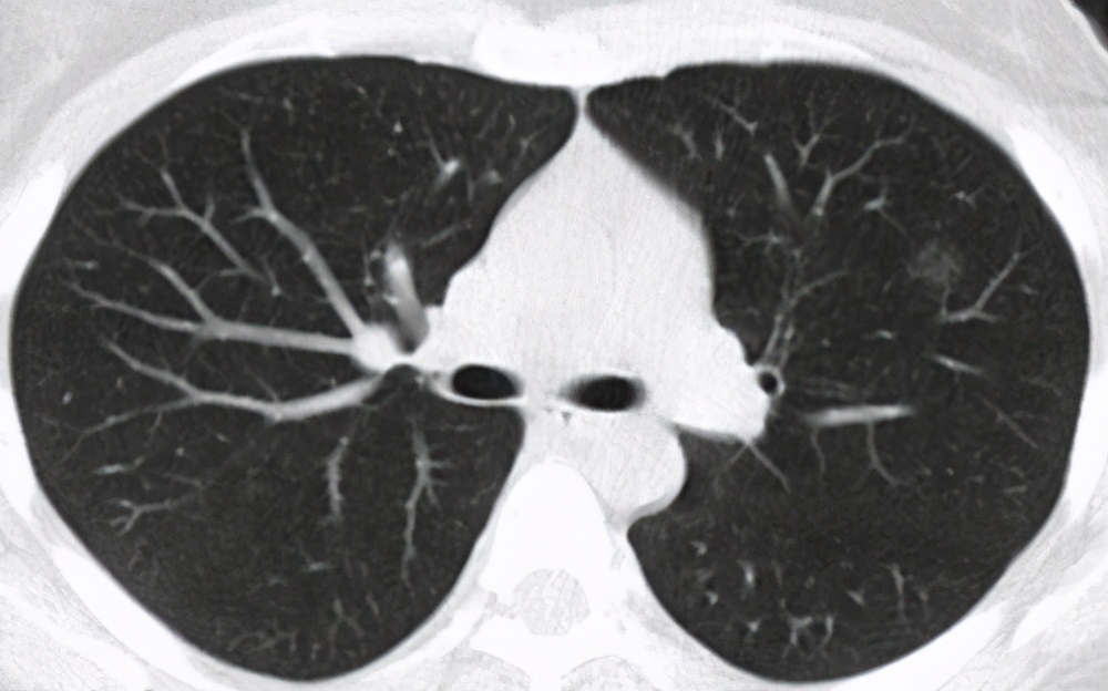
Ground-glass opacities in the lungs are a finding on chest CT scans, typically caused by various inflammations, edema, fibrosis, tumors, etc. They are particularly significant for the early diagnosis of lung cancer.
Due to the impact of this pandemic, many people underwent chest CT scans with the initial intention of screening for the virus. However, this led to the discovery of many early-stage lung cancer cases.
II. Detection of Lung Cancer
Historically in clinical practice, most lung cancer patients we encountered were diagnosed at an advanced stage, as lung cancer often presents no noticeable symptoms in its early phases. In routine health check-ups, chest CT scans are rarely included—usually only chest X-rays are performed. Compared to CT scans, X-rays are not as accurate and cannot detect tiny lesions. Therefore, we often hear from patients that despite undergoing annual check-ups, no abnormalities were ever detected.
The most common symptom of lung cancer in its early stages is coughing, which manifests as paroxysmal, irritating dry cough with little or no phlegm. Some patients may also experience hemoptysis, blood-streaked sputum, difficulty breathing, hoarseness, and other symptoms. Many patients go to the hospital for examination due to these symptoms, leading to the detection of lung cancer.

Additionally, 5%-15% of patients are discovered during routine physical examinations or chest imaging studies without any symptoms present at the time of detection.
One important point to emphasize is that 75% of patients are already at an advanced stage when they first seek medical attention, which is a key reason for the high mortality rate of lung cancer.
III. When is lung cancer screening necessary?
Lung cancer screening should be performed when the following manifestations occur:
1. Abnormal lung findings are detected during physical examination or pulmonary imaging studies;
2. Unexplained irritating cough with poor response to treatment
3. Unexplained pleural effusion;
4. Recurrent hemoptysis or blood-streaked sputum;
5. Recurrent pneumonia in the same location, ineffective or only briefly effective with anti-inflammatory treatment;
6. History of chronic respiratory disease, with recent worsening cough, choking cough, or cough with a high-pitched, metallic sound.
IV. What are the examination methods for lung cancer?
1. Laboratory tests
Tumor marker tests have certain reference value for the diagnosis of lung cancer, such as carcinoembryonic antigen (CEA), neuron-specific enolase (NSE), cytokeratin 19 fragment antigen (CYFRA21-1), progastrin-releasing peptide (ProGRP), squamous cell carcinoma antigen (SCC-Ag), etc.
Tests such as complete blood count and liver and kidney function help assess the patient's overall condition but cannot serve as a basis for diagnosing lung cancer.
2. Imaging Examination
Chest X-ray examination is an important means for early detection of lung cancer, being convenient, simple, and easy to promote; however, its drawback is that the accuracy is not high enough and it cannot display tiny lesions.
CT is currently the most important method for screening lung cancer. It can detect early-stage lung cancer and display the location and size of lesions. Given the current economic and medical levels, it is a relatively convenient, safe, and economical method for lung cancer screening.
PET-CT is currently the most advanced imaging examination method, which can effectively show malignant tumors throughout the body. It is generally used for examining metastatic cancer and malignancies where the primary lesion cannot be determined.
3. Endoscopy
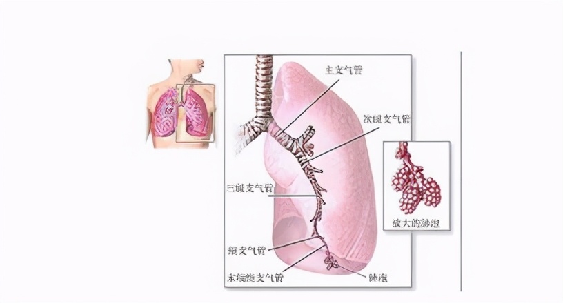
Bronchoscopy is one of the most commonly used diagnostic methods for lung cancer. It allows direct observation of lesions within the bronchi and is suitable for confirming central lung cancer.
Mediastinoscopy is primarily used for diagnosis in patients with mediastinal lymph node metastasis who are not suitable for surgery.
Thoracoscopy is suitable for patients who cannot be diagnosed via bronchoscopy or needle biopsy.
4. Pathological Examination
Fine-needle aspiration is one of the most commonly used diagnostic methods for lung cancer. For patients who cannot be diagnosed via bronchoscopy, fine-needle aspiration can be used for confirmation.
Cytological examination of cells obtained through endoscopy, needle biopsy, pleural fluid, or sputum can provide a preliminary diagnosis.
Biopsy of lung tissue is the gold standard for diagnosing lung cancer.
5. Genetic Testing
Performing genetic testing on lung cancer tissue facilitates the implementation of targeted therapy.